Banner image placeholder
Banner image

In Situ Confocal Raman Microscopy of a Glycolated Polythiophene Mixed Conductor


Journal article


Kabery Rani Sarker, Tahamida Alam Oyshi, Robert Posey, Joshua Tropp, Carol Korzeniewski
Chem. Mater., ASAP Article, 2026


Cite

Cite

APA   Click to copy
Sarker, K. R., Oyshi, T. A., Posey, R., Tropp, J., & Korzeniewski, C. (2026). In Situ Confocal Raman Microscopy of a Glycolated Polythiophene Mixed Conductor. Chem. Mater., ASAP Article. https://doi.org/10.1021/acs.chemmater.6c01121


Chicago/Turabian   Click to copy
Sarker, Kabery Rani, Tahamida Alam Oyshi, Robert Posey, Joshua Tropp, and Carol Korzeniewski. “In Situ Confocal Raman Microscopy of a Glycolated Polythiophene Mixed Conductor.” Chem. Mater. ASAP Article (2026).


MLA   Click to copy
Sarker, Kabery Rani, et al. “In Situ Confocal Raman Microscopy of a Glycolated Polythiophene Mixed Conductor.” Chem. Mater., vol. ASAP Article, 2026, doi:10.1021/acs.chemmater.6c01121.


BibTeX   Click to copy

@article{kabery2026a,
  title = {In Situ Confocal Raman Microscopy of a Glycolated Polythiophene Mixed Conductor},
  year = {2026},
  journal = {Chem. Mater.},
  volume = {ASAP Article},
  doi = {10.1021/acs.chemmater.6c01121},
  author = {Sarker, Kabery Rani and Oyshi, Tahamida Alam and Posey, Robert and Tropp, Joshua and Korzeniewski, Carol}
}
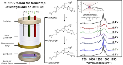
Conjugated polymer-based organic mixed ionic-electronic conductors (OMIECs) undergo complex, coupled changes in electronic structure and ion content as their oxidation state is varied electrochemically. Experimental approaches that can probe these transformations within electrode-supported films under operational conditions are needed to guide molecular design. Here, a confocal Raman microscope equipped with a high numerical aperture (NA) oil-immersion objective is applied as an in situ spectroelectrochemical probe of conjugated polythiophene OMIEC films. Using high-performing glycolated bithiophene–thienothiophene copolymer (PgBTTT) as a model with regioregular poly(3-hexylthiophene) (RR-P3HT) serving as a benchmark, Raman bands associated with neutral, polaronic, and bipolaronic states are identified and tracked as a function of applied potential. Measurements on films ranging from 200 nm to ∼10 μm demonstrate depth-selective in situ probing of OMIEC doping and effects consistent with resistive or transport limitations. Furthermore, this approach enables simultaneous differentiation of backbone and side chain responses, offering a potential handle on the coupled ionic-electronic processes that define OMIEC function. Such observations are not readily accessible with standard benchtop in situ gravimetric or optical absorption spectroelectrochemical techniques. These results establish confocal Raman microscopy as an accessible, sensitive, depth-selective tool for interrogating oxidation-state evolution in situ within OMIECs relevant to bioelectronics, sensing, and energy applications.

Share

Text Only Owlstown
QR Code
Translate to